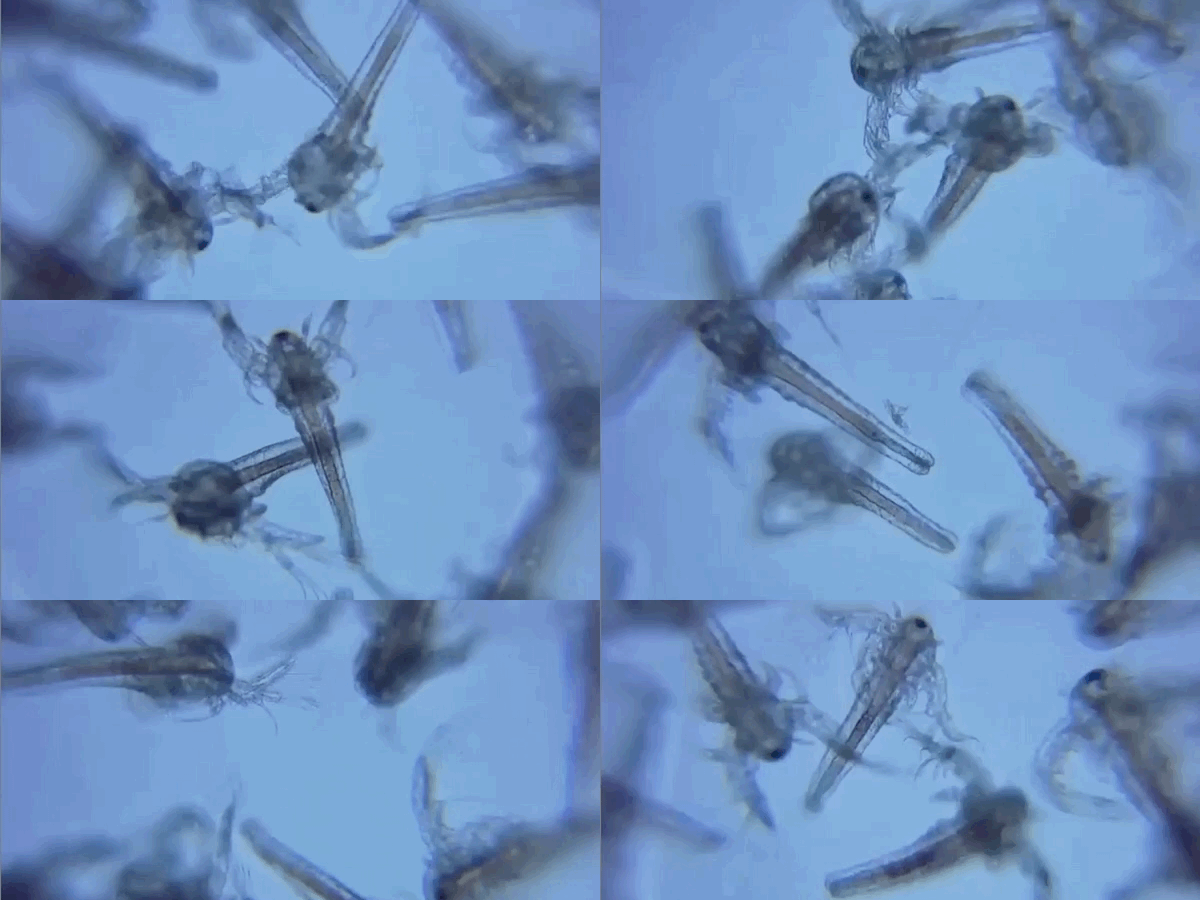

Smartphone microscopy sounds a little like a science-fair stunt: take the same device you use for texts, maps, and photos of suspiciously pretty coffee, clip on a tiny lens, and suddenly you are peeking into a hidden world. But this is not a gimmick. Over the past several years, researchers, teachers, engineers, and citizen scientists have shown that smartphone microscopy can be a genuinely useful way to view cells, fibers, plant structures, parasites, droplets, and other tiny details without hauling around a full lab setup. In some cases, these systems are built for classrooms. In others, they support fieldwork, telemedicine, and point-of-care testing in places where traditional microscopes are expensive, bulky, or simply unavailable.
The big appeal is easy to understand. Smartphones already combine a camera, screen, battery, processing power, storage, and connectivity in one pocket-sized slab. Add a lens, some lighting, and a stable mount, and that slab starts moonlighting as a microscope. It will not replace every benchtop instrument. It will not magically turn your phone into a Nobel Prize machine. But it can make microscopy more affordable, portable, and shareable. That is a pretty good trick for something that also still has your grocery list.
What Is Smartphone Microscopy?
Smartphone microscopy is the use of a smartphone camera, usually paired with an external optical attachment or adapter, to capture magnified images of very small objects. The setup can be as simple as a clip-on macro or ball lens, or as advanced as a custom optical module with fluorescence, darkfield, or confocal imaging capabilities. Some systems attach directly to the rear camera. Others connect the smartphone to a standard laboratory microscope so the phone acts as the camera and display.
In plain English, it is microscopy built around the hardware most people already own. That is why the field matters so much. Instead of asking schools, clinics, or field teams to buy a full imaging station first, smartphone microscopy starts with the camera already in your hand. From there, the add-ons can range from delightfully scrappy to impressively sophisticated.
How Smartphone Microscopy Works
The optics do the heavy lifting
A smartphone camera alone is designed for everyday photography, not for studying onion skin, pollen grains, or red blood cells. To get microscopic detail, the phone needs extra optics. Common approaches include clip-on macro lenses, reversed phone camera lenses, tiny glass spheres, and custom objective systems. These additions shorten the working distance and let the camera focus on objects that are extremely close.
Some research designs use surprisingly elegant tricks. A paper-based Foldscope can create significant magnification with a tiny spherical lens. Other systems use a second phone lens mounted in front of the phone’s existing lens. More advanced versions add filters, UV illumination, or optical paths that allow brightfield, darkfield, or fluorescence imaging. So yes, beneath the humble clip lies a lot of optical wizardry. Tiny wizardry, but still wizardry.
Lighting matters more than people expect
A blurry microscope image is often not a “bad camera” problem. It is a lighting problem. Good smartphone microscopy depends on even illumination, controlled contrast, and a stable sample. Some devices use the phone’s built-in flash. Others rely on ambient light, LEDs, or transmitted light passing through a sample on a slide. Fluorescence systems go further by using specific wavelengths and filters so only selected structures glow.
This is why two smartphone microscopes with similar-looking lenses can produce very different results. If the lighting is uneven, the specimen is too thick, or the sample is wobbling like a caffeinated jellyfish, the image quality drops fast.
Software quietly improves the experience
Software is one of the secret advantages of smartphone microscopy. A phone can autofocus, enlarge the live view on screen, capture images instantly, tag location data, and send files to another person in seconds. In research and medical settings, apps can also assist with counting features, measuring size, or analyzing patterns. That said, smartphone software can also be annoying. Automatic sharpening, color correction, and exposure settings may make a specimen look prettier while making the data less trustworthy. In other words, your phone may be trying to help when what you really need is honesty.
Why Smartphone Microscopy Matters
Traditional microscopes are powerful, but they are not always practical. They can be expensive, fragile, heavy, and difficult to distribute widely. Smartphone microscopy changes the equation by lowering the barrier to entry. It can reduce cost, increase portability, and make it easier to document and share what you see. For schools, that means more students can get hands-on access to microscopy. For field teams, it means bringing imaging to the sample instead of always bringing the sample back to the lab. For remote health or environmental work, it creates new ways to screen, document, and communicate findings.
It also changes the social side of microscopy. A traditional microscope is often a one-person experience: one eye to the eyepiece, one tiny gasp, one triumphant “I found it.” A smartphone microscope turns that into a shared screen, a photo, a video, a group discussion, or a remote consult. That shift matters just as much as the optics.
Real-World Uses of Smartphone Microscopy
Classroom learning and STEM outreach
This is one of the most exciting uses. Smartphone microscopes make it easier to put microscopy in the hands of students who may never have access to a full lab. Teachers can build or distribute low-cost kits, guide students through sample collection, and have them capture images using devices they already know how to use. Instead of treating microscopy like a museum piece behind glass, smartphone microscopy turns it into something active, personal, and portable.
Students can examine salt crystals, flower petals, strands of hair, insect wings, fabrics, sand grains, pollen, paper fibers, and leaf structures. That may sound simple, but simple is often the gateway drug to scientific curiosity. One weird-looking speck becomes a question. One question becomes ten. Suddenly the classroom is full of future biologists and one kid who is definitely going to magnify a potato chip.
Field science and citizen science
Smartphone microscopy works well outside the lab because it travels easily. Citizen scientists can document tiny organisms in water, insects, plant disease symptoms, or surface textures in the field and share images quickly. Researchers have long recognized that portability matters. A device that fits in a pocket or small kit can go places a standard microscope never will, including classrooms, farms, remote clinics, and outdoor survey sites.
Point-of-care diagnostics
In global health and low-resource settings, smartphone microscopy has been studied for malaria, parasitic infections, blood analysis, and other forms of rapid imaging. Some systems are designed to identify parasites or visualize blood-smear features. Others pair microscopy with microfluidic chips, specialized stains, or automated image analysis. The advantage is not just low price. It is the combination of portability, digital capture, and the ability to move information quickly.
This does not mean every phone microscope is clinic-ready. Diagnostics require validation, training, repeatability, and careful sample preparation. But the progress is real, and it is one reason smartphone microscopy has earned serious attention from universities and research labs.
Remote consultation and telemedicine
Because the image is already digital, smartphone microscopy naturally fits remote review. A teacher can project an image for a class. A student can upload a specimen photo for discussion. A field worker can send images to a specialist. A clinic can document a sample and share it for a second opinion. The microscope becomes not just an imaging device, but also a communication device. That is a major shift from the old “trust me, it looked weird in the eyepiece” method of scientific reporting.
What You Can Actually See
With a decent smartphone microscope, you can often see plant cells, pollen, textile fibers, paper fibers, insect anatomy, algae, protozoa, hair structure, skin flakes, and the shapes of many tiny materials. Some well-designed systems can visualize blood cells and other clinically relevant features. Fluorescence versions can highlight structures that would otherwise be difficult to distinguish.
There are limits, though. Smartphone microscopes are not ideal for everything. Viruses are well beyond the reach of ordinary optical smartphone setups. Very fine diagnostic work can demand more resolution, better control, and more reliable sample preparation than a casual clip-on device can provide. So the right mental model is not “a phone can do everything a lab microscope can do.” It is “a phone microscope can do more than most people expect, especially when the setup is thoughtfully designed.”
Common Smartphone Microscopy Setups
Clip-on macro lenses
These are the most beginner-friendly option. They are inexpensive, widely available, and easy to snap on. They are great for larger fine details such as insect parts, fabrics, fingerprints, or plant surfaces. They are not always “true microscopy,” but they are often the easiest first step.
Ball lenses and reversed lenses
These produce stronger magnification and can reveal genuinely microscopic detail. The tradeoff is that they often have a tiny field of view and can be harder to use. Getting the subject centered may feel like trying to park a bus on a postage stamp.
Paper or 3D-printed microscope builds
These are popular in education and DIY communities because they are low-cost and surprisingly capable. Paper-based microscopes and 3D-printed clips can be distributed at scale, customized, and repaired or replaced cheaply.
Phone-to-bench-microscope adapters
This setup uses a conventional microscope for the optics and the smartphone for image capture. It is less portable, but extremely useful for documentation, sharing, and teaching. It is also a smart option when you want the quality of a traditional microscope with the convenience of digital capture.
The Biggest Strengths of Smartphone Microscopy
- Affordability: many systems cost far less than a traditional microscope setup.
- Portability: ideal for classrooms, travel, fieldwork, and home exploration.
- Digital sharing: images and videos can be saved, sent, and discussed instantly.
- Accessibility: lowers the barrier for beginners and resource-limited environments.
- Integration: the camera, screen, battery, processing, and storage are already built in.
The Biggest Limitations
Smartphone microscopy also has real weaknesses, and pretending otherwise would be sloppy. First, the field of view can be small, which makes scanning a specimen slower. Second, image quality may vary from one phone model to another. Third, automatic camera settings can distort color or reduce measurement accuracy. Fourth, many specimens still require careful preparation, and sample prep is often the least glamorous but most important part of the whole process.
Stability is another issue. Even tiny movements can ruin the shot. Focus can be finicky. Illumination can be uneven. And while low-cost builds are fantastic for learning and many practical tasks, they do not erase the need for validated clinical instruments when decisions about diagnosis or treatment are on the line. Smartphone microscopy is powerful, but it is not a hall pass out of rigor.
Tips for Better Smartphone Microscopy Results
- Use a stable stand or rest your setup on a solid surface.
- Control lighting instead of relying on random room illumination.
- Clean the lens, the slide, and the phone camera before every session.
- Use thin, simple specimens when possible; thick samples are harder to image well.
- Lock focus and exposure if your camera app allows it.
- Capture several images rather than trusting the first decent-looking one.
- Take notes on the lens, lighting, and sample so you can reproduce what worked.
Is Smartphone Microscopy Worth It?
Yes, with one important condition: you need to match your expectations to the setup. If you want a compact, affordable, and surprisingly capable tool for education, exploration, field documentation, or certain targeted imaging tasks, smartphone microscopy is absolutely worth it. If you expect every clip-on lens to perform like a high-end research microscope, disappointment will arrive quickly and with dramatic flair.
The smartest way to think about smartphone microscopy is as a bridge. It bridges curiosity and observation, classrooms and labs, remote settings and specialists, low-cost tools and meaningful data. It makes the tiny world more reachable. And that matters, because science often begins not with giant machines, but with the simple act of noticing something small and deciding it deserves a closer look.
Experiences With Smartphone Microscopy: What It Feels Like in Real Life
Using smartphone microscopy for the first time is usually a mix of delight, confusion, and the sudden realization that dust is everywhere. You clip on the lens, open the camera, and assume tiny miracles will appear immediately. Instead, the first few minutes are often a comedy of almosts: too blurry, too dark, too close, too far, wrong angle, mystery thumb in frame. Then, quite suddenly, something snaps into focus. A grain of pollen looks sculpted. A strand of hair resembles a rope. The edge of a leaf becomes a landscape. That little moment is the hook.
In classrooms, the experience tends to become social very quickly. One student finds a useful specimen, and everyone wants to try it. Someone brings in flower petals. Another person discovers that notebook paper is not smooth at all. A teacher projects the live image, and the room turns into a tiny-world theater. What makes smartphone microscopy especially engaging is that it feels familiar. Students already understand the camera interface. They know how to pinch, tap, save, and share. That familiarity lowers the intimidation factor that traditional lab equipment sometimes creates.
For hobbyists and curious adults, the experience is a little different. Smartphone microscopy tends to turn ordinary objects into personal field studies. You start with the obvious things like salt, sugar, cloth, or a feather. Then you move on to stranger choices because the microscope makes everything look like it is hiding a secret. Coffee grounds. Dust from a windowsill. A basil leaf. The wing of a dead insect you found on the porch. The joy is not just in the magnification. It is in the shift in attention. You begin to look at the world as a collection of surfaces, structures, and patterns waiting to be noticed.
In more practical settings, the experience becomes less whimsical and more purposeful. Field teams value the speed. Health workers value portability. Researchers appreciate being able to capture and send images on the same device. But even in serious contexts, the emotional experience is often the same: satisfaction when a lightweight system produces an image that is actually useful. That is the charm of smartphone microscopy. It keeps surprising people by being more capable than it looks.
The frustrations are real, too. Focus can drift. Lighting can be fussy. The field of view can feel tiny. Sometimes you spend more time adjusting the sample than observing it. But that frustration is part of the learning curve, and it teaches an important lesson: microscopy is not just about magnification. It is about preparation, patience, and technique. Once people figure that out, the quality of their results improves dramatically.
In the end, the experience of smartphone microscopy is memorable because it changes how people see ordinary life. It turns the phone from a passive screen into an active scientific tool. It invites curiosity. It rewards patience. And once you have seen the geometric drama inside a grain of salt or the alien architecture of a flower petal on a device you usually use to check the weather, it is hard not to want another look.
Final Thoughts
Smartphone microscopy is not a toy pretending to be science. At its best, it is real science made more portable, affordable, and approachable. Researchers have used it for diagnostics, imaging, and quantitative analysis. Educators use it to make discovery feel immediate. Makers use it to build tools on a budget. Beginners use it to turn everyday objects into tiny adventures. That range is exactly what makes the topic so compelling.
The technology will keep improving as phone cameras, optics, software, and 3D-printed accessories get better. But the most important part is already here: smartphone microscopy gives more people the chance to look closely. And in science, looking closely is where the interesting stuff begins.


