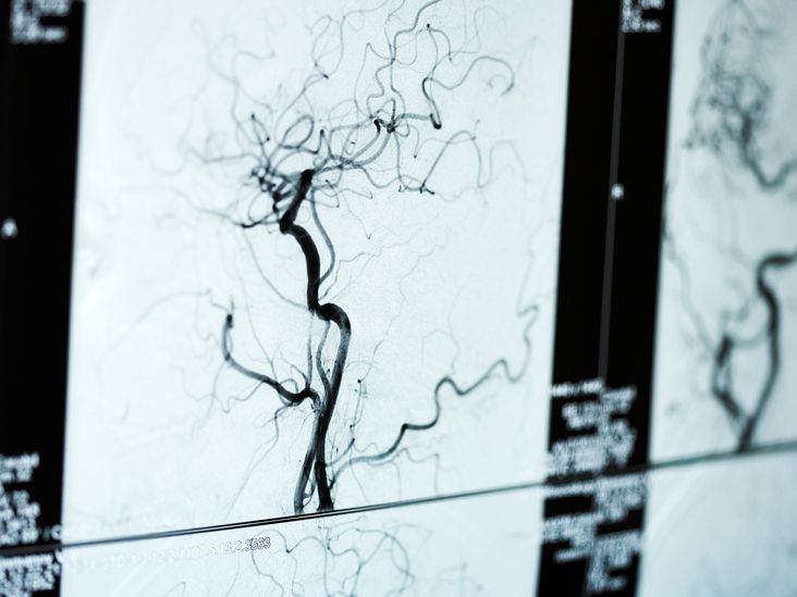

An arteriogram may sound like something a hospital printer spits out when it is having a very serious Monday, but it is actually one of the clearest ways doctors can see what is happening inside your arteries. Also called arterial angiography, this imaging test uses contrast dye and X-ray technology to create detailed pictures of blood vessels. In plain English: it helps your care team find narrow spots, blockages, aneurysms, bleeding, abnormal vessels, and other artery problems that ordinary exams cannot always reveal.
If your doctor has recommended an arteriogram, it does not automatically mean bad news is waiting around the corner wearing a tiny lab coat. Often, the test is ordered to confirm a diagnosis, plan treatment, or guide a procedure such as angioplasty, stent placement, clot treatment, or embolization. This guide explains what an arteriogram is, the main types, how to prepare, what happens during the procedure, what the results may show, and what real-life experience around the test often feels like.
What Is an Arteriogram?
An arteriogram is a medical imaging test used to examine arteries, the blood vessels that carry oxygen-rich blood away from the heart. During a traditional catheter arteriogram, a doctor places a thin, flexible tube called a catheter into an artery, commonly through the wrist, arm, or groin. Contrast dye is then injected through the catheter while X-ray images are taken. The dye makes the arteries visible, creating a moving or still “road map” of blood flow.
The words arteriogram, angiogram, and arteriography are often used together. Technically, an arteriogram focuses on arteries, while angiography can refer to imaging of arteries or veins. In everyday medical conversations, many people use “angiogram” as the broader term. Either way, the goal is the same: to show whether blood is moving normally or running into a traffic jam.
Why Doctors Order an Arteriogram
Doctors may recommend an arteriogram when symptoms, physical exams, ultrasound, CT scans, MRI scans, or blood tests suggest a possible artery problem. The test can help identify the location, severity, and shape of a blood vessel issue. That information matters because treatment choices often depend on tiny details, such as whether a blockage is short or long, whether a vessel is twisted, or whether an aneurysm has a narrow or wide neck.
Common Reasons for an Arteriogram
An arteriogram may be used to evaluate chest pain, leg pain with walking, poor wound healing in the feet, stroke-like symptoms, unexplained bleeding, suspected aneurysms, kidney artery narrowing, tumors with unusual blood supply, or traumatic injury to blood vessels. In some cases, it is performed in an emergency, such as when doctors need to locate active internal bleeding or restore blood flow quickly.
Diagnosis and Treatment Can Happen Together
One major advantage of catheter angiography is that it can be both diagnostic and therapeutic. If the doctor finds a blocked or narrowed artery, they may be able to treat it during the same session with balloon angioplasty, a stent, clot-removal tools, or medicine delivered directly into the vessel. That is medical efficiency at its finest: find the plumbing problem and, when appropriate, fix it before the patient even changes out of the hospital gown.
Main Types of Arteriogram
The type of arteriogram depends on which arteries need to be studied. Although the basic idea is similar, different body areas require different planning, imaging angles, and specialist expertise.
Coronary Arteriogram
A coronary arteriogram looks at the coronary arteries, which supply blood to the heart muscle. It is often performed during cardiac catheterization to check for coronary artery disease. Doctors may order it for chest pain, abnormal stress test results, suspected heart attack, or planning before certain heart procedures. If a significant blockage is found, angioplasty and stent placement may sometimes be performed during the same visit.
Cerebral Arteriogram
A cerebral arteriogram examines arteries in the brain, head, or neck. It may help evaluate aneurysms, arteriovenous malformations, narrowed arteries, blood clots, tumors with rich blood supply, or certain causes of stroke symptoms. Because the brain is involved, this test is handled with special care and detailed monitoring.
Peripheral Arteriogram
A peripheral arteriogram studies arteries in the arms, legs, hands, or feet. It is commonly used in people with peripheral artery disease, especially when symptoms include calf pain while walking, numbness, cold feet, slow-healing wounds, or skin color changes. The results can help doctors decide whether lifestyle treatment, medication, angioplasty, stenting, or surgery is the best next step.
Renal Arteriogram
A renal arteriogram focuses on the arteries that supply the kidneys. It may be used when doctors suspect narrowing of a kidney artery, certain causes of high blood pressure, aneurysms, vascular injury, or abnormal blood flow related to kidney disease. Kidney function is especially important to discuss before this test because contrast dye is processed through the kidneys.
Aortic Arteriogram
An aortic arteriogram evaluates the aorta, the body’s largest artery. Doctors may use it to assess aneurysms, dissections, trauma, or blood flow to major branch vessels. Modern CT angiography often provides excellent aortic images, but catheter angiography may still be useful in selected cases, especially when treatment is planned.
Pulmonary Angiography
Although the pulmonary arteries carry blood from the heart to the lungs rather than oxygen-rich blood to the body, pulmonary angiography is sometimes discussed alongside arteriogram procedures. It may be used to study blood flow in the lungs, though CT pulmonary angiography is now more common for suspected pulmonary embolism.
CT Angiography and MR Angiography
Not every artery-imaging test requires a catheter. CT angiography uses computed tomography and contrast dye through an IV to create detailed 3D images of blood vessels. MR angiography uses magnetic resonance imaging, sometimes with contrast and sometimes without. These tests are less invasive than catheter angiography, but they may not allow immediate treatment during the same exam.
How to Prepare for an Arteriogram
Preparation depends on the type of arteriogram, the body area being studied, your medical history, and whether sedation will be used. Your care team should give specific instructions, and those instructions matter more than random advice from your neighbor, your cousin, or a forum post written in all caps.
Tell Your Doctor About Medications and Allergies
Before the procedure, tell your healthcare team about all prescription medications, over-the-counter drugs, supplements, and allergies. Blood thinners, diabetes medications, kidney-related medicines, and drugs that affect clotting may require special instructions. Never stop a medication on your own unless your doctor tells you to do so.
Discuss Kidney Function and Contrast Dye
Most catheter arteriograms use iodinated contrast dye. For many people, this is safe, but extra caution is needed for patients with reduced kidney function, dehydration, diabetes, heart failure, or a history of contrast reaction. Your doctor may order blood tests before the procedure and may recommend hydration or other precautions.
Fasting and Transportation
You may be asked not to eat or drink for several hours before the test, especially if sedation is planned. Many patients also need someone to drive them home afterward. Even if you feel alert, sedation can make your reflexes and judgment take a little vacation.
What to Bring
Bring a list of medications, insurance information, identification, and any relevant imaging reports. Wear comfortable clothing and leave jewelry at home. If you use glasses, hearing aids, or mobility aids, ask the facility what you can keep with you before and after the test.
What Happens During the Arteriogram Procedure?
Most arteriograms are performed in a hospital, catheterization lab, or interventional radiology suite. You will usually change into a gown, lie on a procedure table, and have monitors placed to track your heart rhythm, blood pressure, and oxygen level.
Step 1: Numbing the Access Site
The doctor cleans the skin and injects local anesthetic to numb the access site. Common access points include the radial artery in the wrist or the femoral artery in the groin. You may feel a brief sting from the numbing medicine, but the goal is to prevent sharp pain during catheter placement.
Step 2: Placing the Catheter
A small puncture is made in the artery, and a thin catheter is guided through the blood vessel using real-time X-ray imaging. The doctor carefully steers the catheter to the area being studied. You should not feel the catheter moving inside the artery, though you may feel pressure at the insertion site.
Step 3: Injecting Contrast Dye
Contrast dye is injected through the catheter while X-ray images are taken. Some people feel warmth, flushing, or a brief strange taste when the dye is injected. That sensation usually passes quickly. It can feel odd, but odd is not the same as dangerous.
Step 4: Taking Images and Possibly Treating a Problem
The care team captures images from different angles. If a treatable problem is found, the doctor may perform an intervention during the same procedure, depending on the reason for the test, your consent, and the clinical situation.
Step 5: Removing the Catheter and Closing the Site
When imaging is complete, the catheter is removed. Pressure, a closure device, or a band may be used to reduce bleeding. If the groin was used, you may need to lie flat for a period of time. If the wrist was used, a compression band may be placed around the wrist.
How Long Does an Arteriogram Take?
The imaging portion may take less than an hour for some patients, but the full visit is usually longer because of preparation, monitoring, recovery, and possible treatment. Complex procedures, emergency cases, or combined diagnostic and therapeutic work can take more time. Many patients go home the same day, but some need overnight observation, especially after heart, brain, or more involved vascular procedures.
What Do Arteriogram Results Show?
Arteriogram results show how blood flows through the arteries and whether the vessels look normal or abnormal. A radiologist, cardiologist, vascular surgeon, neurologist, or other specialist may interpret the images depending on the type of test.
Normal Results
Normal results generally mean that blood appears to flow through the studied arteries without major narrowing, blockage, aneurysm, abnormal connection, or active bleeding. However, “normal” must be interpreted in the context of symptoms. If symptoms continue, your doctor may recommend other tests to look for non-artery causes.
Narrowing or Blockage
One of the most common findings is stenosis, which means narrowing of an artery. A complete blockage may also be seen. These findings can be caused by atherosclerosis, blood clots, inflammation, injury, or other conditions. The report may describe the percentage of narrowing, the length of the blockage, and whether blood is finding alternate routes through collateral vessels.
Aneurysm
An aneurysm is a bulging or ballooning area in a blood vessel wall. Arteriography can help show the size, shape, location, and blood-flow pattern of an aneurysm. This information helps doctors decide whether the aneurysm should be monitored, repaired, or treated with an endovascular technique.
Abnormal Blood Vessel Connections
Some people have abnormal connections between arteries and veins, such as arteriovenous malformations or fistulas. These can occur in the brain, spine, kidneys, limbs, or other areas. An arteriogram can show how blood enters and leaves the abnormal connection, which is essential for treatment planning.
Active Bleeding
In trauma, gastrointestinal bleeding, or post-surgical bleeding, an arteriogram may show contrast leaking from an artery. In selected cases, doctors can treat the bleeding by placing tiny materials through the catheter to block the problem vessel, a process called embolization.
Risks and Possible Side Effects
An arteriogram is generally safe when performed by trained specialists, but every medical procedure has possible risks. Common minor effects include bruising, soreness, or a small lump at the access site. These usually improve with time and basic aftercare.
Less common risks include bleeding, infection, allergic reaction to contrast dye, damage to a blood vessel, blood clots, kidney problems related to contrast, or abnormal heart rhythm. Rare but serious complications may include stroke, heart attack, emergency surgery, or severe bleeding. The exact risk depends on the type of arteriogram, the patient’s health, and whether treatment is performed during the procedure.
When to Call a Doctor After an Arteriogram
Call your healthcare team right away if you have increasing pain, swelling, bleeding that does not stop with pressure, fever, drainage from the access site, numbness, weakness, chest pain, shortness of breath, severe headache, trouble speaking, or a cool or discolored limb. Do not play medical detective at home when symptoms are serious. This is not the moment for “let’s see what happens.”
Recovery After an Arteriogram
After the procedure, nurses monitor your vital signs and check the access site for bleeding. You may be asked to keep your arm or leg still for a while. Drinking fluids may help flush contrast dye from the body, unless your doctor has limited your fluids for another medical reason.
Most patients are told to avoid heavy lifting, strenuous exercise, and soaking in a bath or pool for a short period. Showering is often allowed after a specific time, but instructions vary. Follow your discharge papers carefully, because the wrist and groin do not read minds.
Arteriogram vs. CT Angiogram vs. Ultrasound
A catheter arteriogram offers highly detailed images and allows doctors to treat some problems immediately. CT angiography is less invasive and can provide excellent 3D views, especially for the heart, brain, lungs, aorta, and limbs. MR angiography may be useful when radiation exposure or iodinated contrast is a concern, although it is not suitable for every patient or every device implant. Ultrasound is often used first for peripheral artery disease, carotid artery disease, aneurysm screening, and blood-flow checks because it is widely available and does not use radiation.
The “best” test is not the fanciest one. It is the one that answers the clinical question safely and clearly. Your doctor chooses based on symptoms, urgency, kidney function, allergies, prior imaging, and whether treatment may be needed at the same time.
Questions to Ask Before Your Arteriogram
Before the test, consider asking: Why do I need this arteriogram? Which arteries are being studied? Will this be catheter angiography, CT angiography, or another type? What are the alternatives? Should I stop any medications? How should I prepare if I have diabetes, kidney disease, or a contrast allergy? Could treatment be done during the same procedure? When will I get the results? What symptoms after the test should make me call immediately?
These questions are not annoying. They are practical. Good healthcare works better when patients understand the plan instead of nodding bravely while secretly wondering whether “contrast dye” is something used in printer cartridges.
Experience-Based Insights: What an Arteriogram Can Feel Like in Real Life
Many people feel nervous before an arteriogram, not because they are fragile, but because the idea of a catheter entering an artery sounds like a plot twist nobody requested. In real-world patient experiences, the waiting period before the test is often more stressful than the procedure itself. The room may look high-tech, the staff may use unfamiliar terms, and monitors may beep with the confidence of tiny robots. Knowing what to expect can make the experience feel more manageable.
One common experience is surprise at how organized the process is. Patients are usually checked in, asked repeated safety questions, given an IV, and prepared by nurses who have done this many times. The repeated questions may feel excessive, but they are part of safety checks: confirming your name, allergies, medications, procedure site, and medical history. It is not that the team forgot who you are. It is that healthcare safety loves repetition almost as much as toddlers love asking “why.”
During the procedure, most people report pressure rather than sharp pain after the numbing medicine takes effect. The contrast injection can create a warm sensation that spreads quickly and disappears. Some people feel mildly flushed. Others feel nothing dramatic and wonder whether they missed the big moment. Sedation, when used, may make patients relaxed and drowsy, but many remain awake enough to follow simple instructions such as holding still or taking a breath.
Recovery can feel slow because stillness is part of the job. If the groin was used, lying flat can be uncomfortable, especially for people with back stiffness. If the wrist was used, the compression band may feel tight. Nurses check the pulse, skin color, temperature, and puncture site. Patients sometimes underestimate how tired they may feel afterward. Even when the test goes smoothly, the combination of fasting, stress, sedation, and hospital logistics can leave the body asking for a nap with excellent legal representation.
The emotional side of results also matters. If the arteriogram shows normal arteries, relief may arrive fast, followed by the next question: “Then what caused my symptoms?” If it shows a blockage or aneurysm, the patient may feel overwhelmed. A helpful approach is to ask the doctor to explain the finding in practical terms: where it is, how severe it is, what it means, whether it needs treatment now, and what the next step will be. A diagram or image review can make the explanation much clearer.
Patients who have a smoother experience often do a few simple things well: they bring an accurate medication list, follow fasting instructions, arrange a ride home, ask questions beforehand, and avoid rushing back into normal activities too quickly. They also pay attention to discharge instructions, especially about lifting, driving, bathing, and watching the access site. The test may be high-tech, but recovery still appreciates old-fashioned common sense.
For families and caregivers, the best support is calm, practical help. Drive the patient home, help track instructions, prepare an easy meal, and encourage rest. Avoid turning the evening into a dramatic medical documentary. Most people need reassurance, hydration if allowed, and a comfortable place to relax. If warning symptoms appear, contact the medical team rather than guessing.
The biggest lesson from patient experience is that an arteriogram is not just a test; it is a decision-making tool. It can turn vague symptoms into a clear map, help doctors choose the right treatment, and sometimes fix a problem immediately. That does not make the procedure fun, exactly. Nobody adds “arteriogram” to a vacation bucket list. But with good preparation, clear communication, and careful follow-up, it can be a powerful step toward answers.
Conclusion
An arteriogram is a detailed artery-imaging test that helps doctors see how blood flows through specific blood vessels. It can diagnose narrowing, blockages, aneurysms, abnormal vessel connections, and bleeding. Depending on the situation, it may also guide treatment during the same procedure. While risks exist, careful screening, modern imaging tools, trained specialists, and good aftercare help reduce complications.
If your doctor recommends an arteriogram, the smartest move is to understand the purpose, prepare well, and ask clear questions. The more you know, the less the procedure feels like a mystery and the more it becomes what it really is: a highly detailed map of your body’s vascular roads.


